9 results
Some microsporidia found in certain fishes and insects in eastern Canada
- H. B. Fantham, Annie Porter, L. R. Richardson
-
- Journal:
- Parasitology / Volume 33 / Issue 2 / May 1941
- Published online by Cambridge University Press:
- 06 April 2009, pp. 186-208
-
- Article
- Export citation
-
An account is given of some Microsporidia found in certain fishes and insects in eastern Canada, among which are new species of Nosema, Perezia and Gurleya.
Nosema pimephales n.sp. produces large cysts or tumours in the abdomen of Pimephales promelas, one out of a large number from Lake Guindon, Province of Quebec, being infected.
Nosema branchiale Nemeczek has been observed from a cod, Gadus callarias. The organism is shown to be a Nosema, though the incomplete original description has caused doubts as to whether some other genus was involved. Further details are now given.
Nosema apis Zander and N. bombi Fantham & Porter have been found in Canada in hive-bees and humble-bees respectively, the latter being recorded from a new host, Bombus vagans.
Glugea hertwigi n.var. canadensis is described from Osmerus mordax from Lake Edward.
Perezia aeschnae n.sp. has been found in nymphs of the large dragonfly, Aeschna grandis, collected near Montreal and is described and illustrated.
Perezia legeri Paillot has been observed in larvae of Pieris brassicae.
These appear to be the first records of Perezia from insects in North America, and the zoogeographical distribution of the genus is extended to the Odonata.
Gurleya aeschnae n.sp, is described from a nymph of Aeschna grandis. It is the first species of Gurleya to be described from the Odonata and the first record of the genus from the North American continent.
Thelohania corethrae Schuberg & Rodriguez is described from the larvae of Chaoborus flavicans. Measurements of the parasite, which were lacking in the original description, are now supplied.
Thelohania bracteata from larvae of Simulium bracteatum and larvae and one adult S. venustum and Thelohania fibrata from larvae of Simulium venustum are recorded from places in the Province of Quebec. The need of reinvestigation of the species of Thelohania from various Simulium and the possibility of there being but one species, Thelohania varians Debaisieux, are indicated.
Thelohania legeri Hesse is recorded as a parasite of larvae of Anopheles punctipennis in Quebec Province and of A. gambiae in Zululand. It is probably the most widely distributed species of Thelohania, occurring in Anophelines in Europe, Asia, Africa and North America.
Some general remarks regarding zoogeographical distribution, habitat, host-specificity and possible origin of species and varieties in connexion with Gurleya, Perezia and Thelohania are presented.
The Spirochaetes found in the Crystalline Style of Tapes aureus: A Study in Morphological Variation
- H. B. Fantham
-
- Journal:
- Parasitology / Volume 2 / Issue 4 / December 1909
- Published online by Cambridge University Press:
- 06 April 2009, pp. 392-408
-
- Article
- Export citation
-
One of the earliest known Spirochaetes from Lamellibranchs was that briefly described by Certes in 1882 from the crystalline style of the oyster. Certes named the parasite Trypanosoma balbianii. In 1891 the same worker recorded the occurrence of the organism in two species of Tapes, namely T. decussatus and T. pullaster. Laveran and Mesnil in 1901 showed that Tr. balbianii probably belonged to the Spirochaetes. The organism is now known as Spirochaeta balbianii.
Observations on Theileria parva, the Parasite of East Coast Fever of Cattle
- George H. F. Nuttall, H. B. Fantham, Annie Porter
-
- Journal:
- Parasitology / Volume 2 / Issue 4 / December 1909
- Published online by Cambridge University Press:
- 06 April 2009, pp. 325-340
-
- Article
- Export citation
-
Although a good deal has been written about East Coast Fever in cattle, the literature relating thereto contains very little direct information regarding the parasite which stands in causal relation to the disease. Robert Koch (1898), who was the first to observe the parasite in cases of East Coast Fever occurring in German East Africa, regarded it as but a variety of Piroplasma bovis (= bigeminum) and described the disease as Texas Fever. Theiler (1904) was the first to distinguish clearly East Coast Fever from Redwater. He stated that “the disease has nothing to do with Texas Fever or Redwater; it is a new disease due to a parasite different to the one found in Texas Fever.” Koch (1903—1904), who gave the disease its distinctive name, reached the same conclusions as Theiler. The investigations of Theiler (1904) established the following facts: Cattle which are immune to Redwater are susceptible to East Coast Fever. East Coast Fever is not communicable by blood inoculations (30 experiments, wherein 5 to 2000 c.c. of East Coast Fever blood were inoculated). He noted the absence of haemoglobinuria in the majority of animals affected with East Coast Fever, its presence in the majority of the animals affected with Redwater. He found that in most cases of East Coast Fever, there was no appreciable decrease in the number of red blood corpuscles, this being in marked contrast to what is observed in Redwater. Theiler noted that cattle might harbour both the parasites of Redwater (P. bovis) and those of East Coast Fever (bacillary forms = T. parva). The former generally appeared in the blood “only towards the end of the fever reaction in East Coast Fever,” being previously latent in the animals which had been “salted” against Redwater. He distinguished “two groups of piroplasmosis,” the inoculable (Redwater, canine and equine piroplasmosis) and the uninoculable (East Coast Fever) by injection of infected blood. The parasites in the latter are much smaller than in the former. He named the parasites of East Coast Fever Piroplasma parvum. Theiler distinguished the parasite of East Coast Fever from P. bovis because of the frequent occurrence of bacillary forms and the minute size of the parasite, but he nevertheless retained the new parasite in the genus Piroplasma.
Theileria parva, the Parasite of East Coast Fever in Cattle: Observations on Stained Preparations
- George H. F. Nuttall, H. B. Fantham
-
- Journal:
- Parasitology / Volume 3 / Issue 2 / July 1910
- Published online by Cambridge University Press:
- 06 April 2009, pp. 117-129
-
- Article
- Export citation
-
In an earlier paper (Nuttall, Fantham and Porter, 1909, Parasitology vol II. pp. 325—340), we recorded our observations on living Theileria parva as seen in the peripheral blood of two cows which succumbed to East Coast Fever1. We now propose to describe our studies upon the parasite in stained preparations made from the animals' blood during the course of the disease and from their organs shortly after death. We shall confine our attention to the types of parasites encountered within red blood corpuscles or to corresponding types which may be occasionally encountered free in the plasma. The subject of “Koch's blue bodies” will receive attention at a later date.
Some Haematozoa observed in vertebrates in eastern Canada
- H. B. Fantham, Annie Porter, L. R. Richardson
-
- Journal:
- Parasitology / Volume 34 / Issue 2 / July 1942
- Published online by Cambridge University Press:
- 06 April 2009, pp. 199-226
-
- Article
- Export citation
-
Some new species of Mastigophora and Sporozoa and a Microfilaria, parasitic in fish, frogs and toads in eastern Canada, are described and illustrated.
Trypanosoma percae n.var. canadensis from the yellow perch, Perca flavescens, is monomorphic, smaller, with a shorter, broader body and shorter free flagellum than Trypanosoma percae from European Perca fluviatilis. Trypanosoma myoxocephali n.sp. from the longhorn sculpin, Myoxocephalus octodecimspinosus, is small, with a narrow undulating membrane. It is the first trypanosome to be recorded from Myoxocephalus. Trypanosoma lavalia n.sp. from Bufo americanus is delicate, with a prominent undulating membrane with two myonemes. Trypanosoma gaumontis n.sp. from Bufo americanus is small, crook-like, with undulating membrane with deep folds and one myoneme very-near the bordering flagellum. There is no free flagellum. Trypanosoma montrealis n.sp. from Bufo americanus is long and sinuous, with very short free flagellum and without chromatoid granules in the cytoplasm.
Trypanosoma rotatorium (Mayer) is described from Rana catesbiana from eight sites, R. clamitans from three sites, and R. pipiens from three sites in Quebec Province. Trypanosoma inopinatum Ed. & Et. Sergent is recorded from Rana catesbiana and R. pipiens, A form similar in dimensions and morphology except for a much shorter free flagellum has been found in R. catesbiana from St Eustache.
Spirochaeta manitoui n.sp. is described from the blood of Rana catesbiana from Lake Manitou.
Lankesterella canadensis n.sp. from Rana catesbiana from Montreal and Lake Manitou is described and illustrated. Intracorpuscular forms have polar vacuoles, schizogony occurs in endothelial cells of the blood vessels of the liver and kidney, and multinucleate oocysts have been observed.
Haemogregarina myoxocephali n.sp. from the longhorn sculpin, Myoxocephalus octo-decimspinosus, and Haemogregarina urophysis n.sp. from the white hake, Urophysis tenuis, are described. Two other haemogregarines sens lat. from species of Rana and Bufoamericanus are described, Haemogregarina aeglifini Henry is recorded from a new host, Gadus callarias from off Labrador, Haemogregarina platessae Lebailly from Pseudopleuronectes americanus and Haemogregarina bigemina Laveran & Mesnil from the eel pout, Zoarces angularis, the hosts being new.
Plasmodium catesbiana n.sp. from Rana catesbiana from the Back River, Montreal, is a small species, with ‘signet ring’ and amoeboid trophozoites, schizonts with up to eight merozoites clustered round coarse pigment granules and gametocytes showing slight sexual dimorphism. Plasmodium bufonis n.sp. occurs in Bufo americanus from Laval des Rapides. The schizonts are large, oval, and give rise to eight merozoites arranged en barillet, the pigment usually being at one pole. Possibly micro- and macromerozoites may be produced. Macrogametocytes are oval and deep-staining; microgametocytes are vermicular. Both have pigment.
A haemosporidian, different from but probably allied to Haemogregarina bigemina, is described from the black sea bass, Centropristis striatus.
Haemoproteus laurentiae n.sp. occurs in Bufo americanus from the banks of the St Lawrence at Montreal South and at Lake Manitou. Its pigment is characteristic, consisting of rod-like or bacilliform distributed granules. Haemoproteus lavalia n.sp. from Bufo americanus, from Laval des Rapides, has pigment restricted to curved bands of rounded to somewhat angular granules. Uninucleate parasites have been observed in extremely few endothelial cells of the lungs. Haemoproteus lanoraiea n.sp. from Bufo americanus, from Lanoraie, has very pale yellow to light brown pigment granules, densely crowded at one pole and forming a compact granular cap. Oval parasites have been found in endothelial cells of the lungs.
Leucocytozoon salvelini n.sp., parasitic in the leucocytes of the speckled trout, Salve-linus fontinalis, is described.
Dactylosoma salvelini n.sp., parasitic in Salvelinus fontinalis from a Laurentian stream, is the second species of Dactylosoma to be described and the third to be notified from fish. It has fan- to wedge-shaped schizonts producing eight merozoites. Its gametocytes are not markedly different from one another in shape but differ in cytoplasmic staining and in nuclear structure. Dactylosoma sylvatica n.sp., parasitic in Rana sylvatica from near Quebec City, produces eight merozoites and has oval and reniform gametocytes.
The morphology of a Microfilaria from Rana sylvatica is described.
The effects of the Protozoa on the host cells are described for each organism, and some differential leucocyte counts for comparable normal and parasitized Amphibia are given in the concluding section.
Some more myxosporidia observed in Canadian fishes
- H. B. Fantham, Annie Porter, L. R. Richardson
-
- Journal:
- Parasitology / Volume 32 / Issue 3 / August 1940
- Published online by Cambridge University Press:
- 06 April 2009, pp. 333-353
-
- Article
- Export citation
-
Seven species of marine fishes and three of fresh or brackish water Canadian fishes have yielded new or known Myxosporidia. The marine fishes were from St Andrews, New Brunswick and Halifax, Nova Scotia, places 250–275 miles apart by sea, and from the Labrador coast. The brackish and freshwater fishes were obtained in Montreal and the Salmon River, Nova Scotia.
Descriptions and illustrations are given of the morphology of five new species of Myxosporidia: Ceratomyxa urophysis, Myxoproteus myoxocephali, Myxidium myoxocephali, Myxosoma diaphana and Henneguya amiae.
Chloromyxum leydigi Mingazzini, C. granulosum Davis, Myxidium lieberkuhni Bütschli, Myxidium gadi Georgévitch and Zschokkella hildae Auerbach are recorded from new piscine hosts in Canada.
Some effects of the Myxosporidian infections, both macroscopic and microscopic, on their hosts are considered. Comparison of the effects of similar infections of Myxosporidia on allied fishes in Normandy and Eastern Canada has shown a general similarity between them.
The economic importance of some Myxosporidian infections of the edible fish examined is shown by parasitism of the cod, haddock and sculpin. Amia calva is also used for food in Montreal. The possibility of edible fishes acquiring Myxosporidian infections by way of their food is noted, with reference to the host-specificity of the Myxosporidia of Fundulus heteroclitus described by Bond.
The influence of environment on and powers of adaptation to different habitats exhibited by certain Myxosporidia is discussed.
Some remarks are made on the zoogeographical distribution of certain Myxosporidia and their piscine hosts.
The Schizogregarines: A Review and a New Classification
- H. B. Fantham
-
- Journal:
- Parasitology / Volume 1 / Issue 4 / December 1908
- Published online by Cambridge University Press:
- 06 April 2009, pp. 369-412
-
- Article
- Export citation
-
1. The term Schizogregarinae Léger (1900) is the name now given to a sub-order of the Gregarinida, the remaining members of which are known as the Eugregarinae. The Schizogregarines were formerly known as Amoebosporidia Aimé Schneider (1884), a name given in misapprehension of the character of the cytoplasmic processes, fixative in function, present in the genus Ophryocystis. Two species of Ophryocystis (0. buetschlii and 0. francisci) were the only members of this sub-order known before 1900.
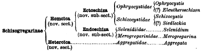
2. At present the sub-order Schizogregarinae contains five families: Ophryocystidae, Schizocystidae, Selenidiidae, Merogregarinidae, and Aggregatidae.
3. All these organisms show well-marked schizogonic stages in their life-history, and—with the possible exception of the Aggregatidae—follow after the Eugregarinae in their methods of sporogony.
4. In Ophryocystis and Schizocystis the schizogony is extracellular, that is, these forms are ectoschizous. The life-cycle of the former is shown in Fig. 1. In these parasites the number of the nuclei in the schizont increases simultaneously with its volume.
5. In Selenidium and Merogregarina the schizogony is intracellular, in other words these forms are endoschizous. The life-cycle of the former is illustrated in Fig. 3. In these forms the schizont is uninucleate during its growth, only becoming multinucleate at the end of the growing period.
6. Ophryocystis forms only one sporocyst, a fact which has been emphasised by Léger and Duboscq (1908), by the placing of the Ophryocystidae in a special section, the Monospora. However, this apparent peculiarity is easily explained by a process of reduction and degeneration having taken place, affecting with one exception all the gametes formed from each gametocyte. There is good morphological evidence in support of this explanation (see p. 382, and Fig. 4, D).
7. Figures of the interesting form Schizocystis gregarinoides (Léger, 1900) are not yet published, but a paper dealing with this organism is promised by Prof. Léger at an early date.
8. Aggregata differs from other Schizogregarines in that its schizogony takes place in one host (crab), while its sporogony occurs in another (Cephalopod mollusc). In this respect Aggregata resembles the Haemosporidia. The schizogonic phases in Crabs were formerly regarded as belonging to a gymnosporous Gregarine, Aggregata Frenzel, while the sporogonic phases were considered to belong to a Coccidian, Eucoccidium (Benedenia) in cuttlefishes and Octopus. Regarding this, Léger and Duboscq (1908, p. 102) write “…Aggregata, avec un changement de cycle coïncidant avec un changement d'hôte, c'est à dire qui soient à la fois digénétiques et hétéroïques.”
9. It is evident therefore that the Aggregatidae stand apart. On this account, I suggest a division of the Schizogregarinae into two sections, termed respectively, the Homoïica (to include the first four families discussed in this paper, wherein schizogony and sporogony take place in the same host) and the Heteroïca (for the Aggregatidae).
10. Among the Homoïca we have extracellular schizogony (ectoschizous forms) in the Ophryocystidae and Schizocystidae, and intracellular schizogony (endoschizous forms) in the Selenidiidae and Merogregarinidae. This difference is not merely superficial, it requires to be emphasised, and for this reason I would divide the section Homoïca into two sub-sections, termed respectively Ectoschiza and Endoschiza.
11. The classification of the Schizogregarines, which I would propose, is as follows:
12. Much further research is needed on the life-cycles of the Endoschiza, especially among the Selenidiidae, which occur so frequently in the Annelida. Sporogonic stages are at present unknown in Eleutheroschizon and Siedleckia.
13. In connection with the Aggregatidae, and to a less extent with the Selenidiidae, stress is laid upon the necessity of carefully distinguishing between “coelomic” and “gut” parasites. (See pp. 397 and 387.)
14. The Schizogregarinae form a most interesting link between the Eugregarinae and the Coccidiidea.
On the Natural Occurrence of Herpetomonads (Leptomonads) in Mice
- H. B. Fantham, Annie Porter
-
- Journal:
- Parasitology / Volume 8 / Issue 1 / June 1915
- Published online by Cambridge University Press:
- 06 April 2009, pp. 128-132
-
- Article
- Export citation
-
While engaged in experimental researches on the introduction of insect flagellates, belonging to the genera Herpetomonas and Crithidia, into vertebrates, we have recalled to mind some former observations of ours made in 1909. When examining the blood of mice artificially infected with the spirochaetes of relapsing fever in the Quick Laboratory, Cambridge, we observed flagellates in the peripheral blood of the mice. These flagellates, which were scanty, were studied in fresh preparations by us and drawings of the organism were made with the camera lucida. Some of these drawings are now reproduced, together with others made by us subsequently. The flagellates were also observed by Professor Nuttall and his laboratory assistant, Mr B. G. Clarke, and we were unanimously agreed that the flagellates belonged to the genus Herpetomonas, or Leptomonas as some have since preferred to call it. The significance of the presence of herpetomonads in the blood of mice was very puzzling at the time, and pressure of other work prevented the subject being followed up. During 1911 and 1912, these flagellate organisms have been seen by all of us, but they were always evanescent and could not be found in fixed and stained preparations. The parasites were seen on different occasions in three mice altogether.
Some Myxosporidia found in certain freshwater fishes water fishes in Quebec province, Canada1
- H. B. Fantham, Annie Porter, L. R. Richardson
-
- Journal:
- Parasitology / Volume 31 / Issue 1 / April 1939
- Published online by Cambridge University Press:
- 06 April 2009, pp. 1-77
-
- Article
- Export citation
-
The Province of Quebec provides a magnificent field for fresh-water faunistic and ecological research on account of its numerous rivers and lakes. Investigations in ecology and ecological parasitology by the authors have revealed a rich fauna in the areas described herein.
Examinations of fourteen species of fresh-water fish have produced twenty-eight species of Myxosporidia, of which twenty-five are new and three have been already described. The existence of races or strains or varieties of some of the forms is demonstrated.
Descriptions and illustrations are given of twenty-eight Myxosporidia found by the authors in fresh-water fishes of Quebec Province. The new species include: Sphaerospora notropis, Myxidium percae, Zschokkella salvelini, Myxosoma notropis, M. commersonii, M. orbitalis, M. pfrille, M. media, M. ellipticoides, M. parellipticoides, Myxobolus poecilichthidis, M. dentium, M. percae, M. rhinichthidis, M. notropis, M. catostomi, M. couesii, M. hybo-rhynchi, M. subcircularis, M. transversalis, M. grandis, Henneguya fontinalis, H. fontinalis var. notropis, H. percae, H. salmonis and H. esocis. Myxosoma catostomi Kudo, Myxobolus conspicuus Kudo and M. ovoidalis Fantham are described from Canadian fishes. Details are given of a series of races, strains or varieties of Thelohanellus notatus Mavor, of Myxobolus catostomi, of M. notropis and of Henneguya fontinalis from other than the original host fish.
The pathology of the myxosporidian infections from the many different situations in the hosts is discussed. Unusual sites of infection are the palatine teeth of Esox maskinongy with Myxobolus dentium and the iris of the eye of Couesius plumbeus with Myxobolus couesii.
In connexion with the economic importance of the Myxosporidia found in Quebec fishes, the destruction of young fish, alteration of the character of the fauna dependent on the same, human food supply, possible epizootics in hatcheries and possible cross-infection of game fishes and other fishes of economic importance are considered. The immediate danger is not great; it is the future that may be imperilled.